1. 引言
急性视网膜坏死综合征(Acute Retinal Necrosis Syndrome (ARN))是一种罕见的病毒感染性葡萄膜炎综合征,表现为以伴有坏死性视网膜炎的全葡萄膜炎,可能会并发视网膜脱离、血管闭塞、视神经病变、玻璃体炎和其它导致视力下降的并发症 [1] 。如果没有准确诊断和治疗,可能会导致毁灭性的视觉后果 [2] 。ARN是由疱疹家族的多个成员引起的,包括水痘带状疱疹病毒(VZV)、单纯疱疹病毒1型和2型(HSV-1, HSV-2)、巨细胞病毒(CMV),以及罕见的E-B病毒(EBV) [3] [4] 。ARN常发生于免疫功能正常的患者,典型病例可以根据临床表现作出诊断,但早期病例及非典型病例的诊断存在困难 [5] ,闫焱等报道 [6] 其误诊率达70%以上,多数患者被误诊为虹膜睫状体炎、全葡萄膜炎、眼内炎,部分患者甚至被误诊为青光眼、视网膜脱离、玻璃体积血等,确诊时,疾病已发展至中晚期。聚合酶链反应检测提供了一种快速、灵敏的方法来确定急性视网膜坏死综合征的病毒病因,为及时准确诊断和治疗提供依据 [7] [8] 。ARN治疗的目的是尽快控制眼部感染,清除病毒,防止并发症,避免视觉损伤。尽管手术、激光和抗病毒治疗取得了进展,ARN的视力预后仍然很差,视网膜脱离率可高达80% [9] [10] 。本文报道二例ARN患者,通过聚合酶链反应检测房水,明确是水痘带状疱疹病毒感染,并采用玻璃体切割术进行了手术治疗,得到了较好的恢复。
2. 病例报告
病例一
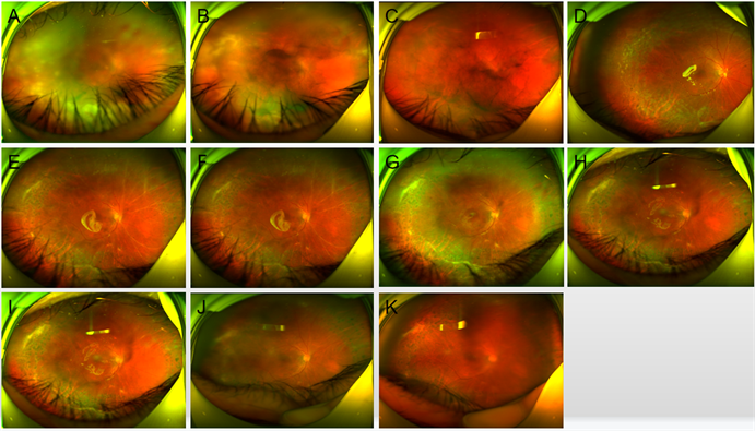
患者,男,56岁,因右眼眼红、胀痛伴视物模糊5天于2021年1月24日第一次入院。体查:Vod指数/20,Vos1.0。右眼结膜混合性充血,角膜内皮见大量羊脂状KP,前房中深,房水混浊,Tyndall征(+),虹膜纹理清,虹膜后粘连,瞳孔形欠圆,直径约4 mm大,对光反射无,晶状体混浊,晶体前区见色素沉着,玻璃体混浊,眼底窥不清。左眼结膜无充血,角膜透明,前房中深,Tyndall征(−),虹膜纹理清,瞳孔圆,直径约3 mm大,对光反射存,晶状体密度增高,玻璃体轻度混浊,眼底:视盘边界清,色红,C/D ≈ 0.3,A/V ≈ 2:3,网膜平伏,黄斑中心凹反光暗(见图1)。诊断为:1) 右眼葡萄膜炎;2) 双眼老年性白内障。诊疗经过:入院后予以完善相关检查,予以地塞米松注射液抗炎、血栓通改善微循环、阿昔洛韦抗病毒等对症支持治疗。2021年2月1日出院情况:Vod:0.15 os1.0;NCT:od13.0 mmHg,os15 mmHg;右眼结膜混合性充血(+),角膜内皮可见KP,前房中深,虹膜纹理欠清,瞳孔药源性散大,直径约5 mm大,对光反射迟钝,晶状体前区可见色素,晶状体密度增高,玻璃体轻度混浊,眼底窥不清。

Figure 1. Case 1: Wide-angle fundus images of the right eye. (A) On January 24, 2021, vitreous opacity, retinal vasculitis and extensive yellow-white necrosis were seen in the retina; (B) On February 1, 2021, the fundus of the left eye showed that the boundary of the optic disc was unclear, the retinal vessels had white line-like changes, and extensive yellow-white necrotic foci were visible in the retina; (C) On March 1, 2021, the vitreous opacity was reduced and the retina was in place; (D) On March 15, 2021, the fundus of the eye after surgery showed that the vitreous cavity was transparent with silicone oil, the retina was flat, and the retina showed laser spots and scars; (E) Fundus map of right eye on March 22, 2021; (F) On April 19, 2021, the right eye fundus picture showed silicone oil in place and retinal scar; (G) On June 1, 2021, silicone oil was in place, retinal scar; (H) on June 23, 2021, refractive stroma opacity, silicone oil was in place, retinal scar; (1) On July 5, 2021, the fundus picture of the right eye after cataract extraction and intraocular lens implantation and silicone oil extraction showed that the refractive stroma was transparent, the retina was in place, and the retinal scar; (J) Retinal scar was seen on July 19, 2021; (K) On July 28, 2021, inflammation subsided and retinal scar
图1. 病例1右眼广角眼底图像。(A) 2021年1月24日见玻璃体混浊,视网膜血管炎改变,视网膜可见广泛黄白色坏死灶;(B) 2021年2月1日左眼眼底图,图中见视盘边界不清,视网膜血管曾白线样改变,视网膜可见广泛黄白色坏死灶;(C) 2021年3月1日见玻璃体混浊减轻,视网膜在位;(D) 2021年3月15日术后眼底图,图中见玻璃体腔硅油透明,视网膜平覆,视网膜见激光斑及瘢痕;(E) 2021年3月22日右眼眼底图;(F) 2021年4月19日右眼眼底图,图中见硅油在位,视网膜瘢痕;(G) 2021年6月1日见硅油在位,视网膜瘢痕;(H) 2021年6月23日见屈光间质混浊,硅油在位,视网膜瘢痕;(I) 2021年7月5日右眼白内障摘除人工晶状体植入硅油取出术后眼底图,图中见屈光间质透明,视网膜在位,视网膜瘢痕;(J) 2021年7月19日见视网膜瘢痕;(K) 2021年7月28日见炎症消退,视网膜瘢痕
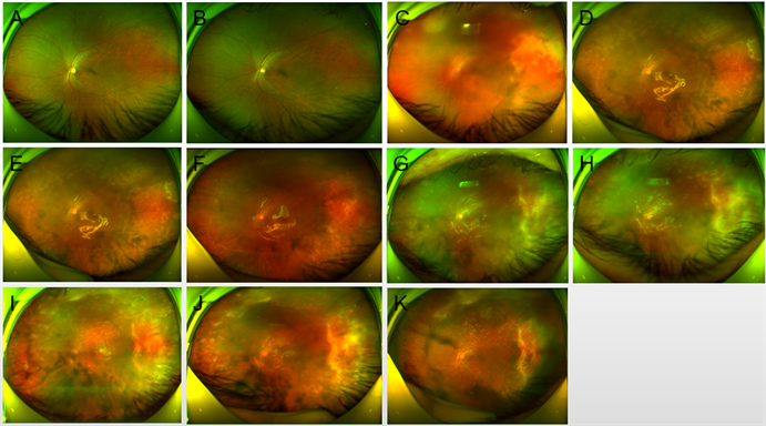
患者因左眼红、视力下降2天,右眼红、视力下降2月2021年2月28日第二次入院。体征:Vod:0.2,os:0.1。矫正:ou:加镜无助。NCT:od12.0 mmHg,os14.3 mmHg。右眼结膜混合性充血,角膜内皮可见KP,前房中深,Tyndalll征(−),虹膜纹理清,部分后粘连,形状不规则,直径约3 mm大,对光反射迟钝,晶状体混浊,玻璃体轻度混浊,眼底窥不清。左眼结膜混合性充血,角膜透明,前房中深,Tyndalll征(−),虹膜纹理清,部分后粘连,形状不规则,直径约3 mm大,对光反射迟钝,晶状体混浊,玻璃体混浊,眼底窥不清(见图1、图2)。取房水进行PCR检查。诊断为:1) 双眼急性视网膜坏死综合征;2) 双眼老年性白内障。诊疗经过:入院后予以完善相关检查,予以地塞米松注射液抗炎、血栓通改善微循环、更昔洛韦抗病毒等对症支持治疗。2021年3月5日行左眼玻璃体切除术 + 视网膜激光 + 硅油填充术,右眼玻璃体腔注射更昔洛韦注射液,2021年3月9日行右眼玻璃体切除术 + 视网膜激光 + 硅油填充术,3月12日行双眼玻璃体腔注射更昔洛韦注射液。2021年3月15日出院时情况:Vou:指数/40,NCT od12.3 mmHg,os11.3 mmHg;双眼结膜混合性充血(+),角膜内皮少许KP,前房中深,虹膜纹理欠清,瞳孔药源性散大,直径约5 mm大,对光反射迟钝,晶状体前囊可见色素,晶状体混浊,玻璃体腔内硅油填充,眼底窥不清,双眼视盘边界清,视色淡红,C/D = 0.3,A:V = 2:3,视网膜平伏,其上可见激光斑,黄斑中心凹反光暗。
患者于2021年6月24日行右眼硅油取出 + 白内障超声乳化人工晶状体植入术;2021年7月20日行左眼硅油取出+白内障超声乳化人工晶状体植入术。2021年7月20日出院时情况:Vod:0.25,矫正:+0.25/−0.75 × 145→0.3−;Vos:0.15−,矫正:+0.50/−3.25 × 35→0.15+。双眼人工晶体在位,玻璃体腔内液体清亮,眼底见视盘边界清,色淡红,C/D约0.3,网膜平伏,周边网膜可见陈旧性激光斑及白色坏死网膜病灶,黄斑中心凹反光暗(见图1、图2)。

Figure 2. Case 1: Wide-angle fundus images of the left eye. (A) On January 24, 2021, the fundus was normal; (B) The fundus was normal on February 1, 2021; (C) On March 1, 2021, vitreous opacity, retinal vasculitis and extensive yellow-white necrosis were seen in the retina; (D) On March 15, 2021, the vitreous cavity was transparent with silicone oil, and retinal laser scar and scar were found after surgery; (E) On March 22, 2021, the retina was in place; (F) On April 19, 2021, silicone oil was in place with retinal scar; (G) On June 1, 2021, the refractive stroma was turbid, silicone oil was in place, and retinal scar was seen; (H) Left eye fundus map on June 23, 2021; (1) Left eye fundus map on July 5, 2021; (J) On July 19, 2021, refractive stroma opacity, silicone oil in place, and retinal scar were seen; (K) On July 28, 2021, the fundus of the left eye after cataract extraction and intraocular lens implantation and silicone oil extraction showed that the retina was in place and retinal scar
图2. 病例1左眼广角眼底图像。(A) 2021年1月24日见眼底正常;(B) 2021年2月1日见眼底正常;(C) 2021年3月1日见玻璃体混浊,视网膜血管炎改变,视网膜可见广泛黄白色坏死灶;(D) 2021年3月15日术后见玻璃体腔硅油透明,视网膜激光瘢及瘢痕;(E) 2021年3月22日见视网膜在位;(F) 2021年4月19日见硅油在位,视网膜瘢痕;(G) 2021年6月1日见屈光间质混浊,硅油在位,视网膜瘢痕;(H) 2021年6月23日左眼眼底图;(I) 2021年7月5日左眼眼底图;(J) 2021年7月19日见屈光间质混浊,硅油在位,视网膜瘢痕;(K) 2021年7月28日左眼白内障摘除人工晶状体植入硅油取出术后眼底图,图中见视网膜在位,视网膜瘢痕
病例二
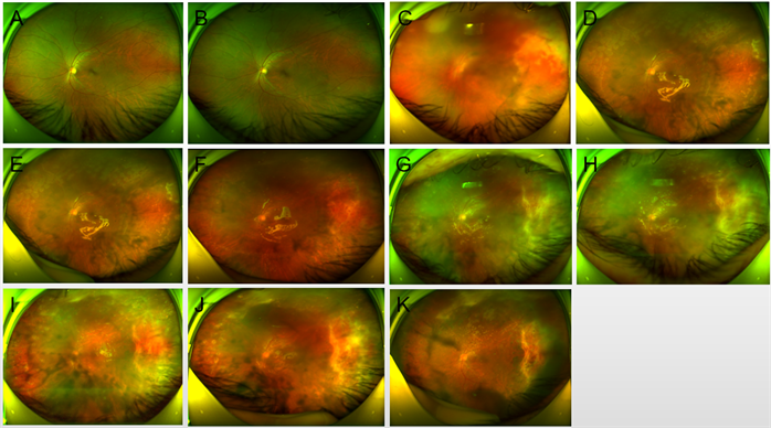
患者,女,57岁,因左眼视力下降1月余于2022年9月26日入院。体查:Vod:0.5,Vos:手动/30 cm,NCTod17.0 mmHg,os13.5 mmHg。右眼结膜无充血,角膜透明,前房中深,Tyndall征(−),虹膜纹理清,瞳孔形圆,直径约3mm大,对光反应可,晶状体混浊,玻璃体轻度混浊,眼底见视盘边界清,色红,C/D ≈ 0.3,A/V ≈ 2/3,视网膜呈豹纹状改变,黄斑中心凹光反射暗。左眼结膜无充血,角膜下方可见陈旧性KP,角膜透明,前房中深,Tyndall征(−),虹膜纹理清,瞳孔形圆,直径约5 mm大,对光反应迟钝,晶状体混浊,玻璃体中度混浊,眼底见视盘窥视欠清,视网膜呈豹纹状改变,视网膜血管可见白线样改变,视网膜可见广泛黄白色坏死灶,已累及黄斑区。B超检查示左眼玻璃体中度混浊。眼底广角照相示左眼视网膜血管可见白线样改变,视网膜可见广泛黄白色坏死灶(见图3)。取房水进行PCR检查。诊断为:1) 左眼视网膜坏死综合征;2) 双眼老年性白内障。于2022年09月27日行左眼玻璃体腔注射更昔洛韦;2022年9月30日行左眼玻璃体切割 + 视网膜激光 + 硅油填充 + 玻璃体注射更昔洛韦术。2022年10月4日出院情况:Vod:0.5,Vos:手动/30 cm,矫正:od:+1.00/−1.25 × 75→0.8,os:+7.50/−1.25 × 90→指数/50 cm;NCT:od15.0 mmHg,os12.0 mmHg;左眼球结膜充血(+),缝线在位,伤口对合可,角膜透明,前房中深,Tyndall(−),瞳孔形圆,约3 mm,晶状体混浊,玻璃体腔内硅油填充,眼底见视盘边界清,视网膜呈豹纹状改变,视网膜血管可见白线样改变,视网膜可见广泛黄白色坏死灶,已累及黄斑区,网膜上可见激光斑。

Figure 3. Case 2: Wide-angle fundus images of the both eyes. (A) On September 26, 2021, the fundus of the right eye was normal; (B) On September 26, 2021, the fundus of the left eye showed that the boundary of the optic disc was unclear, the retinal vessels had white line-like changes, and extensive yellow-white necrotic foci were seen in the retina, which had involved the macular area; (C) On October 4, 2021, the fundus of the left eye after surgery showed that the vitreous cavity was transparent with silicone oil, the retina was flat and the retinal vessels had white line-like changes, and extensive yellow-white necrotic foci were visible in the retina; (D) On October 10, 2021, the left eye fundus picture showed retinal scar; (E) On December 12, 2021, the left eye fundus picture showed local macular detachment; (F) On January 17, 2023, the left eye fundus picture showed silicone oil in place and retinal scar
图3. 病例2广角眼底图像。(A) 2021年9月26日右眼眼底图,图中见眼底正常;(B) 2021年9月26日左眼眼底图,图中见视盘边界不清,视网膜血管呈白线样改变,视网膜可见广泛黄白色坏死灶,已累及黄斑区;(C) 2021年10月4日左眼术后眼底图,图中见玻璃体腔硅油透明,视网膜平覆视网膜血管曾白线样改变,视网膜可见广泛黄白色坏死灶;(D) 2021年10月10日左眼眼底图,图中见视网膜瘢痕;(E) 2021年12月12日左眼眼底图,图中见黄斑局部脱离;(F) 2023年1月17日左眼眼底图,图中见硅油在位,视网膜瘢痕
3. 结果
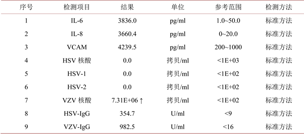
1) 病例1房水PCR检测结果显示水痘带状疱疹病毒(VZV)核酸阳性,IL-6及IL-8升高,可确诊为VZV感染(见图4)。

Figure 4. The PCR results of aqueous humor in case 1 showed that varicella zoster virus (VZV) nucleic acid was positive, and IL-6 and IL-8 were increased
图4. 病例1房水PCR检测结果显示水痘带状疱疹病毒(VZV)核酸阳性,IL-6及IL-8升高
2) 病例2房水PCR检测结果显示水痘带状疱疹病毒(VZV)核酸阳性,可确诊为VZV感染(见图5)。

Figure 5. The PCR results of aqueous humor in case 1 showed that varicella zoster virus (VZV) nucleic acid was positive
图5. 病例2房水PCR检测结果显示水痘带状疱疹病毒(VZV)核酸阳性
4. 讨论
ARN由Urayama等人于1971年首次描述,它具有双峰年龄分布,大约在20多岁和50多岁时达到发病高峰,没有明确的种族关联 [11] 。ARN发病可能是隐匿性的,或者表现为飞蚊症、视力模糊或周边视野缺损。眼外部检查可发现有弥漫性巩膜外层炎、巩膜炎或眼眶炎性疾病。轻度至中度的眼前节细胞反应是常见的,有些病例可出现眼压升高 [12] 。ARN的特征是影响眼底动脉和静脉的视网膜血管炎,表现为血管阻塞。典型病例存在周边视网膜坏死,并且整个周边视网膜可能被累及。玻璃体炎和视盘水肿是ARN的常见特征。ARN可有一个快速发展的过程,通常在发病时只累及一只眼睛。如果不进行治疗,高达70%的患者会出现对侧眼ARN [13] 。对侧受累通常发生在几个月内,但也可能发生在几年后。孔源性视网膜脱离是ARN视力丧失的主要原因,发生率约为50%~80%,玻璃体牵引和增生性玻璃体视网膜病变是导致的主要原因 [14] 。本研究二例病例均为50多岁患者,符合老年发病高峰年龄特征。
ARN的诊断通常是根据临床表现进行的。美国葡萄膜炎学会建立了以下诊断ARN的诊断标准:a)周边视网膜坏死灶;b) 在没有抗病毒治疗的情况下快速发展;c) 闭塞性视网膜血管炎;d) 显著的玻璃体炎和/或前房炎症 [15] [16] 。ARN最常见的病因是由VZV引起的,其次是HSV-1和HSV-2;巨细胞病毒及E-B病毒是ARN的罕见原因 [17] 。典型病例或晚期病例容易诊断,不需要鉴定病毒;但ARN诊断在早期阶段仍然很困难。广角眼底照相能够清楚地记录周边视网膜病变,因为它在单个图像中捕获200˚视角,有利于ARN的早期诊断。红色激光的强穿透力允许广角眼底照相成像穿透混浊的屈光介质 [18] [19] 。即使在患有严重玻璃体炎的患者中,也能够识别坏死的视网膜病变,帮助做出正确的诊断和治疗决策。本研究中,作者利用广角眼底照相较清晰的全程记录了二例患者的病情变化。除此之外,在临床实践中通常寻求通过PCR进行病毒鉴定。PCR是一种高度特异和敏感的试验,可用于检测房水或玻璃体液中的病毒。本研究中,作者对二例患者进行了房水PCR检测,病毒鉴定为VZV,为及时正确诊断提供帮助,也为正确治疗提供依据。在病例不同时间段进行PCR检测,还可以观察眼内病毒量的变化,为长期治疗提供帮助。
静脉注射阿昔洛韦是目前治疗活动性ARN的首选药物。首先用10天的高剂量静脉注射阿昔洛韦进行治疗,然后用口服阿昔洛韦治疗14周。出现复发性病变的患者可能需要长期口服维持治疗 [20] [21] 。更昔洛韦和膦甲酸钠是可供选择的静脉注射药物。玻璃体内注射更昔洛韦或膦甲酸钠可用于治疗受全身性药物毒性限制的个体 [22] [23] 。一些研究者建议抗凝治疗ARN的视网膜血管阻塞患者 [24] [25] 。全身性皮质类固醇有助于控制和限制ARN相关的严重炎症造成的损害 [19] [26] 。有文献建议尽早进行预防性激光屏障治疗,最好在前2周内,以降低视网膜脱离的发生率 [4] [27] 。急进型ARN后伴有多发性视网膜裂孔的孔源性和牵拉性视网膜脱离,玻璃体切除联合硅油填充术是实现视网膜复位的必要手段 [28] 。本研究中,二例患者均采用玻璃体切除联合硅油填充术进行手术治疗,取得了较好的效果。良好的视觉效果可能是由于及时的诊断,适当的和早期的并发症治疗。但本研究只是个案报道,不具有普遍性,尚需要进一步研究来总结经验。
利益冲突
所有作者均声明不存在利益冲突。
基金项目
郴州市科技局项目(lcyl2021117)。
NOTES
*通讯作者。

